AMP, ADP, ATP Nucleotides Separated by HPLC - AppNote
July 19, 2013
/
/
/
/
/
Date: 19-JULY-2013 Last Updated: 31-AUGUST-2026
Separation of AMP, ADP, ATP by HPLC
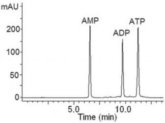
The figure shows a separation of three energy nucleotides. All three are baseline separated in order of increasing polarity as is expected when HILIC chromatography is used.It is worth noting that the retention of nucleotides increased as the buffer concentration was increased (data not shown). 16.0mM concentration of the buffer in Solvents A and B was the maximum concentration still compatible with MS detection.
Peaks:
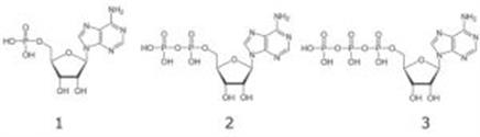
1. AMP – Adenosine 5’-monophosphate
2. ADP – Adenosine 5’-diphosphate
3. ATP – Adenosine 5’-triphosphate
Column: Cogent UDA™, 4 µm, 100 Å
Catalog No.: 40031-05P-2
Dimensions: 2.1 x 50 mm
Mobile Phase:
-- A: DI Water / 16.0 mM Ammonium Formate
-- B: 90% Acetonitrile / 10% DI Water / 16.0 mM Ammonium Formate
Gradient:
| Time (minutes) | %B |
|---|---|
| 0 | 95 |
| 0.5 | 95 |
| 10 | 70 |
| 15 | 30 |
| 20 | 30 |
| 20.1 | 95 |
Post Time: 3 minutes
Injection vol.: 1 μL
Flow rate: 0.4 mL / minute
Detection: UV @ 254 nm
Sample Preparation: Stock Solution: 1 mg / mL solutions in DI Water. Samples were diluted 1:10 into 50% Acetonitrile / 50% DI Water mixture. Before injection, samples were filtered through a 0.45µm Nylon Syringe Filter.
t 0 : 0.7 minutes
Note: The ratio of the Adenine Nucleotides (Adenosine ATP/ADP/AMP) is measured to indicate cell energy status or cell apoptosis/death, or ischemia in a tissue.